IELTS Reading Sample # Imaging Live Tissue
IMAGING LIVE TISSUE
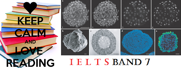
Human breast cancer sample in situ: proteins (green), DNA (magenta), and fat (yellow)PURDUE UNIVERSITY, CHIEN-SHENG LIAOA type of imaging that can capture the activity of proteins, lipids, nucleic acids, and other molecules in some living tissues without the need for fluorescent (the emission of radiation, especially of visible light, by a substance during exposure to external radiation, as light or x-rays.)labels has been in the works in the last decade. But while this technique, called in vivo vibrational (the oscillating, reciprocating, or other periodic motion of a rigid or elastic body or medium forced from a position or state of equilibrium)spectroscopic (an optical device for producing and observing a spectrum of light or radiation from any source, consisting essentially of a slit through which the radiation passes, a collimating lens, and an Amici prism.) imaging (the use of computerized axial tomography,sonography, or other specialized techniques and instruments to obtain pictures of the interior of the body, especially those including soft tissues.), can be used to visualize (to recall or form mental images or pictures)tissues without the need for fluorescent labels, it has still been too slow to be practical for most research (to search or search for again)and clinical applications.
love reading
Now, researchers (A researcher is someone who conducts research, i.e., an organized and systematic investigation into something. Scientists are often described as researchers.)at Purdue University in Indiana have made two major improvements (the action of improving or being improved)to the approach, making it fast enough to be used in real-time and allowing imaging of not just transparent but also thicker, turbid living tissues. The results are published today (October 30) in Science Advances.
“This is a very innovative ( featuring new methods)approach,” said Wei Min of the department of chemistry at Columbia University in New York City who was not involved in the study. “And the instrumentation (measuring instruments regarded collectively)the authors built is quite impressive (evoking admiration through size, quality, or skill; grand, imposing, or awesome).”
“This is good progress toward making this technique (a way of carrying out a particular task, especially the execution or performance of an artistic work or a scientific procedure)more practical,” said bioengineering (the use of artificial tissues, organs, or organ components to replace damaged or absent body parts)professor Stephen Boppart, who develops novel imaging modalities (a particular mode in which something exists or is experienced or expressed)at the University of Illinois and was not involved in the work. “The authors have made the acquisition (the learning or developing of a skill, habit, or quality)faster, allowing image collection in vivo and in highly photon scattering (the process in which electromagnetic radiation or particles are deflected or diffused)tissues.”
While fluorescence microscopy requires labeling a cellular component with a fluorophore, the appeal of in vivo vibrational spectroscopic ( the study of the interaction between matter and electromagnetic radiation. Historically, spectroscopy originated through the study of visible light dispersed according to its wavelength, by a prism)imaging is the ability to produce images that include most of the endogenous (having an internal cause or origin)molecules within tissues or cells without the need to label any cellular components. The original technique sends light through a sample, exciting the molecules in the sample to vibrate at distinct frequencies, which are then registered (enter or record on an official list or directory)as a spectrum or a pattern of peaks. For each pixel, a spectrum (a band of colours, as seen in a rainbow, produced by separation of the components of light by their different degrees of refraction according to wavelength)of frequencies (the rate at which something occurs over a particular period of time or in a given sample)is created and an image is compiled (produce (a list or book) by assembling information collected from other sources)by merging (combine or cause to combine to form a single entity)all of the spectra. A spectrometer (an apparatus used for recording and measuring spectra, especially as a method of analysis)collects the well-directed light that goes through the same, and separates it into its individual wavelengths (a person’s ideas and way of thinking, especially as it affects their ability to communicate with others)while excluding scattered photons—components of light—that decrease the resolution of the light’s spectrum. This method is limited to use for transparent (allowing light to pass through so that objects behind can be distinctly seen.)and single-cell layer biological samples because nontransparent (not able to be seen through; opaque)samples, such as live tissue, scatter too many photons, resulting in poor resolution (the quality of being determined or resolute).
Source : http://www.the-scientist.com/?articles.view/articleNo/44382/title/Imaging-Live-Tissue-Without-Fluorescence/